
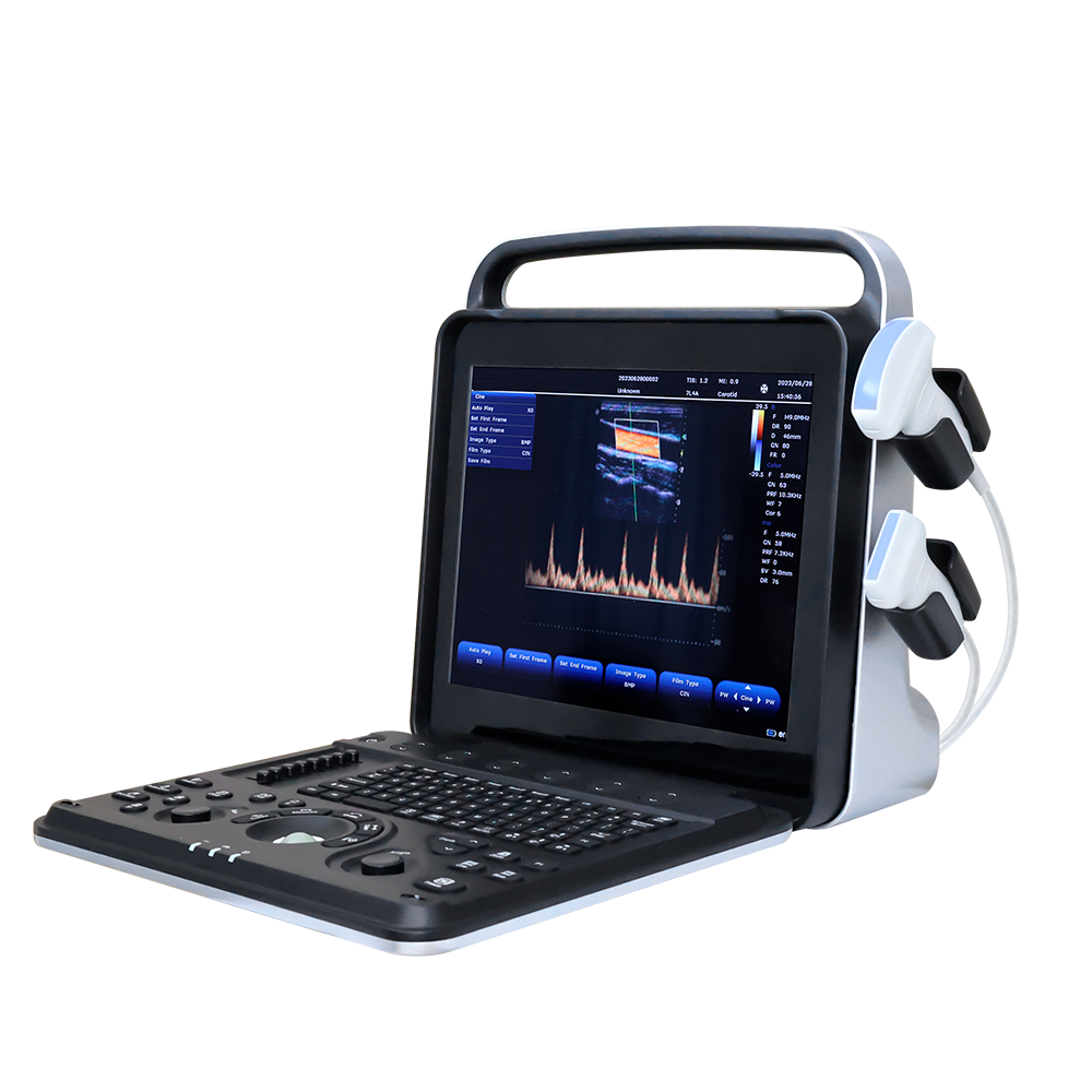
Design highlights:
1. 15 inch medical LCD, full digital 128 elements, 64 channels;
2. Built-in 500 GB hard disk for data storage;
3. Graphics and text management system to enter and classification search medical records;
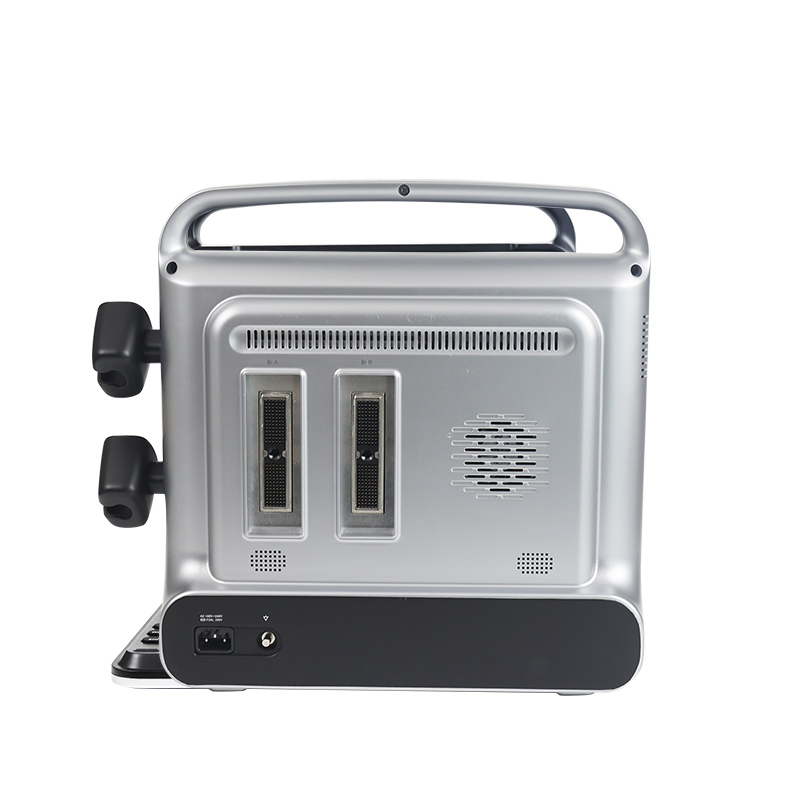
4. Double probe interface, can be used with two probes at the same time;
5. Built-in 18650 lithium battery pack, meet the needs of daily power off use;
6. Special measurement data package for different organs;
7. Images and pathology reports can be exported.
Input / output signal:
1. Input: mquipped with digital signal interface;
2. Output: VGA, s-video,USB, audio interface, network interface;
3. Connectivity: medical digital imaging and communications DICOM3.0 interface components;
4. Support network real-time transmission: can real-time transmission of user data to the server;
5. Image management and recording device: 500G hard disk Ultrasonic image;
6. Archiving and medical record management function: complete
the storage management and playback storage of patient static image and dynamic image in the host computer.
Probe Specifications:
1. 2.0-10MHz V¬ariable frequency, frequency range 2.0-10MHz;
2. 5 kinds of frequencies of each probe, variable fundamental and harmonic frequency;
3. Abdomen: 2.5-6.0MHz;
4. Superficial:5.0-10MHz;
5. Cardiac:2.0-3.5MHz;
6. Puncture Guidance: probe puncture guide is optional, puncture line and Angle are adjustable;
7. Transvaginal: 5.0-9MHZ.
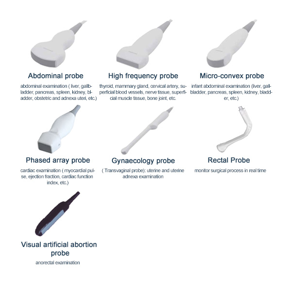
Optional Probes:
1. Abdominal probe: abdominal examination ( liver, gallbladder, pancreas, spleen, kidney, bladder, obstetric and adnexa uteri, etc.);
2. High frequency probe: thyroid, mammary gland, cervical artery, superficial blood vessels, nerve tissue, superficial muscle tissue, bone joint, etc.;
3.Micro-convex probe: infant abdominal examination (liver, gallbladder, pancreas, spleen, kidney, bladder, etc.);
4. Phased array probe: cardiac examination ( myocardial pulse, ejection fraction, cardiac function index, etc.);
5. Gynaecology probe (Transvaginal probe): uterine and uterine adnexa examination;
6. Visual artificial abortion probe: monitor surgical process in real time;
7. Rectal Probe: anorectal examination.
Main technical parameters and functions:
linux +ARM+FPGA
Number of physical channels: ≥64
Number of probe array element number: ≥128
15-inch, high resolution, progressive scan, Wide Angle of view
Resolution:1024*768 pixels
Image display area is 640*480
Internal 500GB hard disk for patient database management
Allow storage of patient studies that include images,clips,reports and measurements
Two active universal transducer ports that support standard(curved array, linear array), high-density Probe
156-pin connection
Unique industrial design provides easy access to all transducer ports
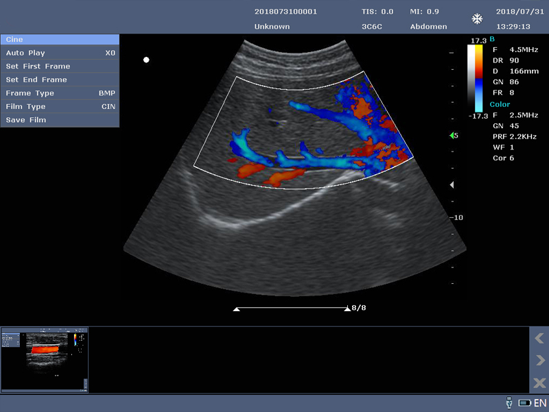
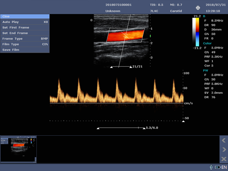
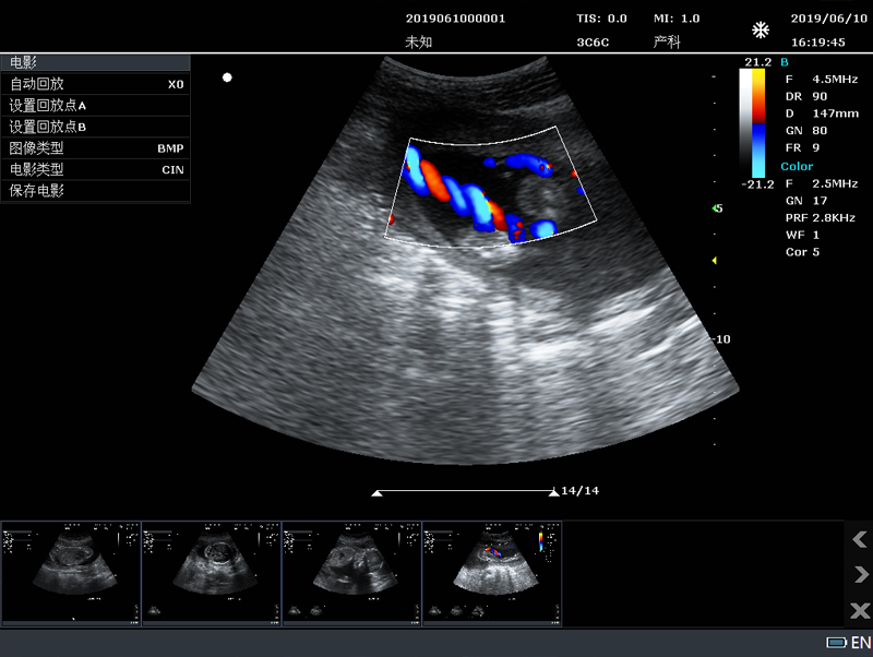
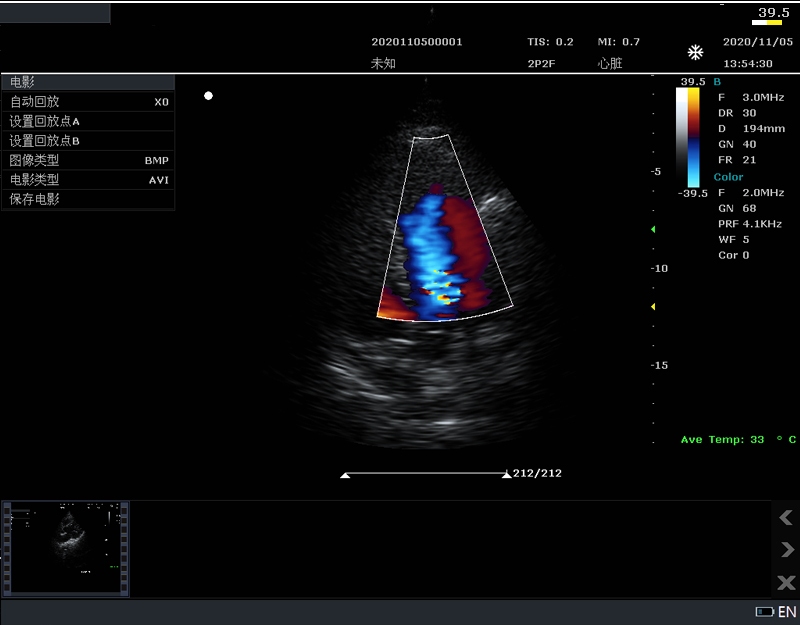
B-mode: Fundamental and Tissue harmonic imaging
Color Flow Mapping (Color)
Power Doppler Imaging (PDI)
PW Doppler
M-mode
B/M:Fundamental wave,≥3; harmonic wave: ≥2
Color/PDI: ≥2
PW: ≥2
B mode: ≥5000 frames
B+Color/B+PDI mode: ≥2500 frames
M、PW: ≥ 190s
available on live, 2B, 4B and reviewed images
up to 10X zoom
format: BMP、JPG、FRM(single image);
CIN、AVI(multiple images)
Support DICOM, conform to DICOM3.0 standard
Built in workstation,support patient data search and browse
Support Chinese, English, Spanish, Can be easily extended to support other languages
Built in large capacity lithium battery, working condition. Continuous working time ≥1.5 hours. Screen provides power display information
Comment、BodyMark、Biopsy、★Lito, etc
2. Ergonomic Design
Frequently used controls centre around the trackball
Control panel is backlighted, waterproof and antisepticised
Two USB port are at the back of the system, which is more convenient for use
3. Exam Modes
Abdomen, Obstetrics, Gynecology, Fetal Heart, Small parts, Urology, Carotid, Thyroid, Breast, Vascular, Kidney, Pediatrics
4. Product configuration
Host(Built-in 500G hard disk)
3C6C convex array probe
7L4C linear array probe
User's Manual
Power cable
USB report printer
B/W or color Video printer
Puncture rack
Trolley
Foot switch
U disk and USB extension line
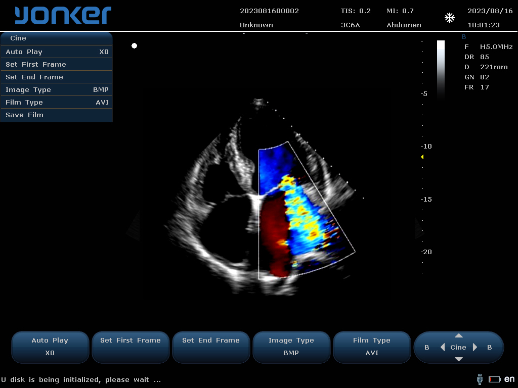
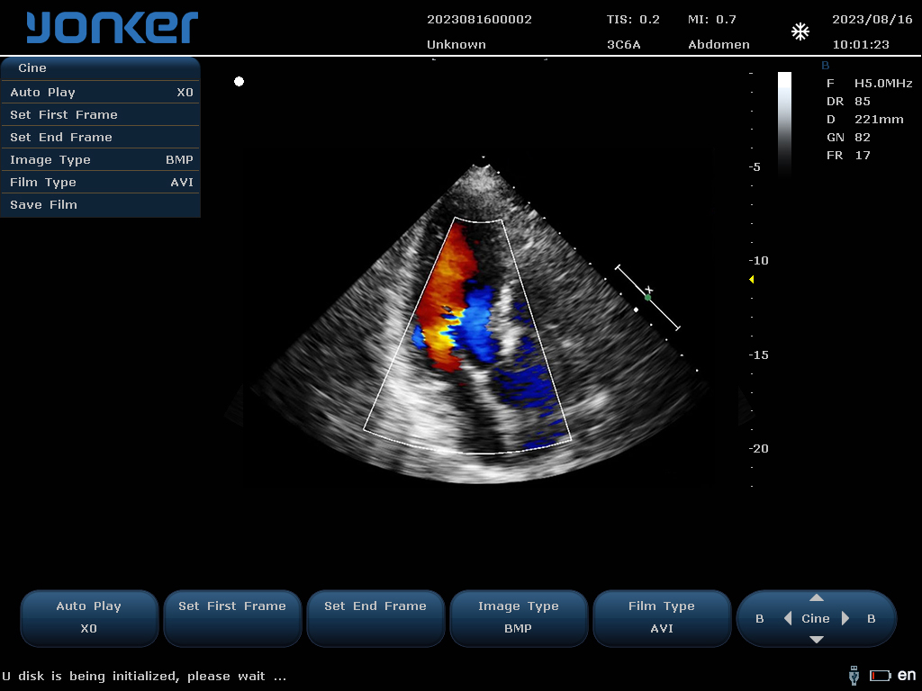
Up to four frequencies in fundamental imaging
Up to two frequencies in Tissue harmonic imaging (probe dependent)
| Dynamic range | 0-100% ,5% step |
| SpeckleReduction | 8 levels(0-7) |
| ScanDensity | H,M,L |
| Gain | 0~100 % ,2% step |
| TGC | eight TGC controls |
| FrameAverage | 8 levels(0-7) |
| LineAverage | 8 levels(0-7) |
| Edge Enhance | 8 levels(0-7) |
| Gray Maps | 15 types(0-14) |
| Pseudocolor Maps | 7 types(0-6) |
| Thermal Index | TIC,TIS,TIB |
| 2B, 4B formats | / |
| Invert (U/D) and transposed (L/R) | / |
| Focus Number | 4 |
| Focus Depth | 16 levels(depth and probe dependent) |
| FOV | 5 levels |
| Image depth up to 35 cm in 0.5~4cm increments (depth dependent) | |
| Phase inversion harmonic imaging technique is available for all probes | |
| Frequency | 2 levels |
| Gain | 0~100% ,2% steps |
| Wall filter | 8 levels(0-7) |
| Sensitivity | H,M,L |
| Flow | H,M,L |
| Packet Size1 | 5 levels(0-4) |
| FrameAverage | 8 levels(0-7) |
| PostProc | 4 levels(0-3) |
| Invert | On/Off |
| Baseline | 7 levels(0-6) |
| Color Maps | 4 levels(0-3) |
| Color/PDI Width | 10%-100%, 10% |
| Color/PDI Height | 0.5-30cm(probe dependent) |
| Color/PDI Center Depth | 1-16cm(probe dependent) |
| Steer | +/-12°,7°(linear probe) |
| Frequency | 2 levels |
| Gain | 0~100% ,2% steps |
| Wall filter | 8 levels(0-7) |
| Sensitivity | H,M,L |
| Flow | H,M,L |
| Packet Size1 | 5 levels(0-4) |
| FrameAverage | 8 levels(0-7) |
| PostProc | 4 levels(0-3) |
| Invert | On/Off |
| Baseline | 7 levels(0-6) |
| PDI Maps | 2 levels(0-1) |
| Color/PDI Width | 10%-100%, 10% |
| Color/PDI Height | 0.5-30cm(probe dependent) |
| Color/PDI Center Depth | 1-16cm(probe dependent) |
| Steer | +/-12°, +/-7°(linear probe) |
| Frequency | 2 levels |
| Sweep speed | 5 levels(0-4) |
| Scale | 16 levels(0-15) (depth and probe dependent) |
| Scale Unit | cm/s,KHz |
| Smooth | 8 levels(0-7) |
| Pseudocolor Maps | 7 types(0-6) |
| Dynamic range | 24-100, 2 step |
| Gain | 0-100%, 2% step |
| Wall filter | 4 levels(0-3) |
| Dynamic range | 24-100, 2 step |
| Gain | 0-100%, 2% step |
| Wall filter | 4 levels(0-3) |
| Angle correction | -89+89,1 step |
| Gate size | 8 levels(0-7mm) |
| Wall filter | 5 levels(0-4) |
| Invert | On/Off |
| Baseline | 7 levels |
| Real-time auto Doppler trace: maximum velocity, mean velocity | |
| Frequency | Up to 3 fundamental and 2 harmonic imaging frequencies |
| Edge enhance | 8 levels(0-7) |
| Dynamic range | 0-100%,step 5% |
| Gain | 0-100,step 2 |
| Gray Maps | 15 levels(0-14) |
| Pseudocolor Maps | 7 (0-6) |
| Sweep speed | 5 levels(0-4) |
★user can press one key to save image parameters in screen
★user can press one key to restore image parameters to default status.
1.Quality Assureance
Strict quality control standards of ISO9001 to ensure the highest quality;
Respond to quality issues within 24 hours, and enjoy 7 days to return.
2.Warranty
All products have a 1 year warranty from our store.
3.Deliver time
Most Goods will be shipped within 72 hours after payment.
4.Three packagings to choose
You have special 3 gift box packaging options for each product.
5.Design Ability
Artwork/Instruction manual/product design according to customer’s requirement.
6.Customized LOGO and Packaging
1. Silk-screen printing logo(Min. order.200 pcs);
2. Laser engraved logo(Min. order.500 pcs);
3. Color box Package/polybag Package(Min. order.200 pcs).