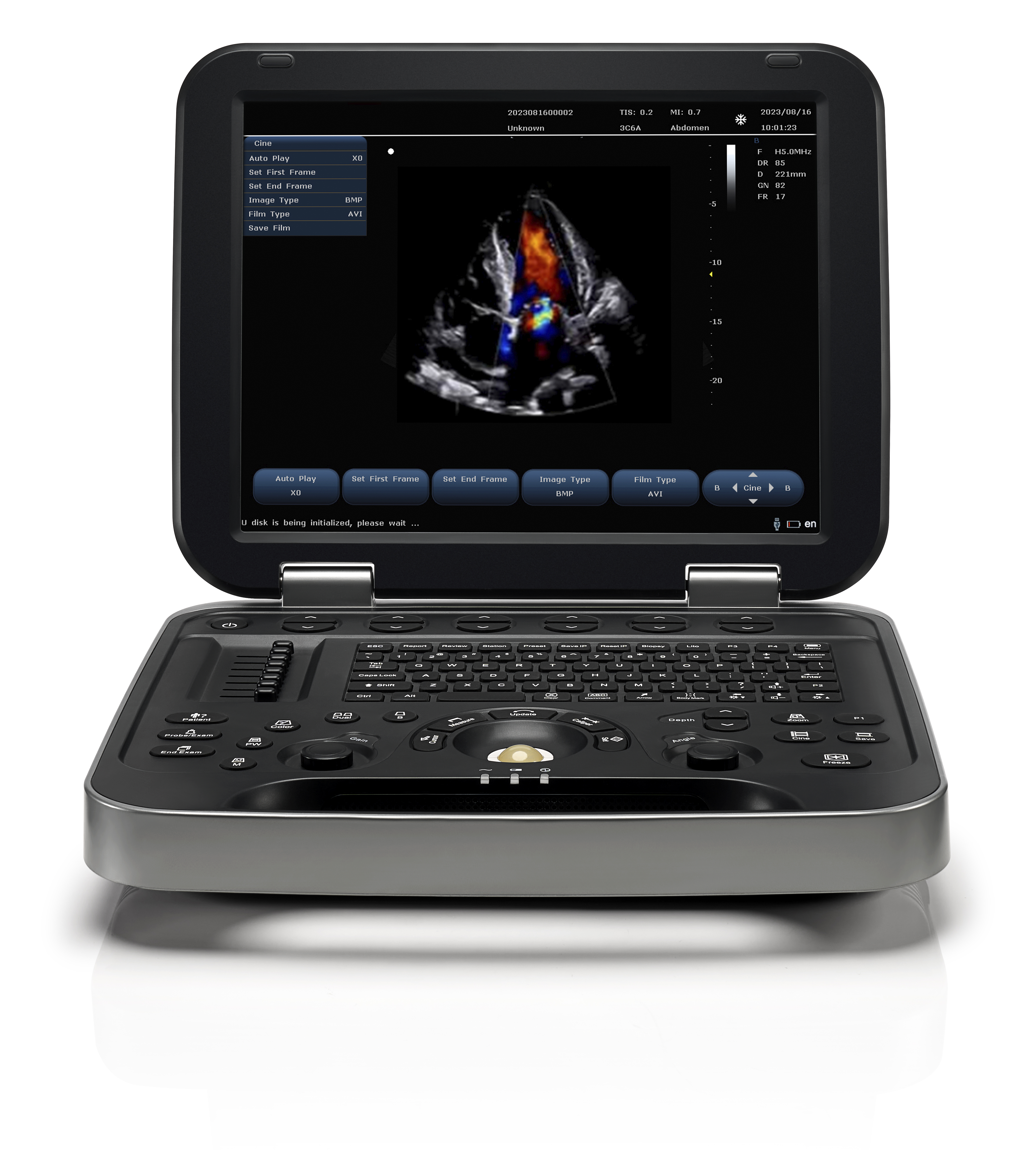
Cardiac Doppler ultrasound is a very effective examination method for clinical diagnosis of heart disease, especially congenital heart disease. Since the 1980s, ultrasound diagnostic technology has begun to develop at an astonishing speed. Like magnetic resonance imaging, CT and isotope scanning, cardiac Doppler ultrasound also has a place in the four major imaging diagnostic technologies in modern medicine.
Cardiac Doppler ultrasound is one of the most important imaging examination technologies in non-invasive cardiac examinations. This examination technology not only has the advantages of being painless, repeatable, harmless and simple, but also has clearer and more accurate examination results than other imaging examinations. After several years of promotion, cardiac Doppler ultrasound has become an indispensable diagnostic tool in modern clinical medicine.
Generally speaking, if the detection result is only mild deficiency, no special treatment is required. If it is moderate or severe heart failure, treatment should be carried out as soon as possible to prevent changes in the patient's heart structure and the possibility of serious complications. In the examination of cardiomyopathy, cardiac Doppler ultrasound can determine the degree of myocardial hypertrophy and cardiac chamber enlargement in patients; for patients with coronary heart disease, cardiac Doppler ultrasound can intuitively show the location of myocardial ischemia, helping clinicians to develop appropriate treatment plans according to the specific conditions of the patients. The main diseases diagnosed by cardiac Doppler ultrasound include aortic lesions (such as lesions such as aortic stenosis), heart valve diseases (such as mitral valve lesions, stenosis, etc.), ventricular diseases, etc.
Cardiac Doppler ultrasound can not only show the distribution of abnormal blood flow in the cardiac cavity, but also reflect the path and direction of cardiac blood flow to a certain extent. It can determine whether the nature of cardiac blood flow is laminar flow, turbulent flow or eddy flow, and can also measure the contour, area, length and specific width of the blood flow beam. Cardiac Doppler ultrasound can directly reflect the relationship between abnormal cardiac structure and abnormal cardiac hemodynamics by displaying blood flow information in a two-dimensional cross-sectional diagram. All children suspected of having congenital heart disease must undergo cardiac Doppler ultrasound examination to determine the specific development of the disease.

Cardiac Doppler ultrasound examination is a very important examination, especially for patients with organic heart disease. Through cardiac Doppler color ultrasound, it is possible to detect whether the subject's heart has structural abnormalities, whether the heart valve has vegetation or other problems. It is also a very reliable reference for evaluating the patient's heart function, examining pericardial disease, and detecting valve function.
The examination of cardiac and cervical vascular Doppler color ultrasound has laid the foundation for our hospital to better serve the surrounding people. Yongkang Medical is a Doppler color ultrasound machine manufacturer with a variety of B-ultrasound color ultrasound machine models. If you find it difficult to choose, Yonkermed Medical can provide detailed color ultrasound machine product information and help recommend several suitable products. It can also take you to experience the operation in person, so that you can buy with confidence.
At Yonkermed, we pride ourselves on providing the best customer service. If there is a specific topic that you are interested in, would like to learn more about, or read about, please feel free to contact us!
If you would like to know the author, please click here
If you would like to contact us, please click here
Sincerely,
The Yonkermed Team
infoyonkermed@yonker.cn
https://www.yonkermed.com/
Post time: Sep-25-2024

