
In addition to the two-dimensional anatomical information obtained by black-and-white ultrasound examination, patients can also use color Doppler blood flow imaging technology in color ultrasound examination to understand the blood flow signal filling distribution of the renal artery, main renal artery, segmental artery, interlobar artery, and arcuate artery of the kidney.
If the blood flow filling of one kidney is significantly reduced or even disappears in the local or entire kidney during the examination, it can be determined that the kidney has renal artery embolism. The color Doppler technology can be used to determine which renal artery is embolized, and even the degree and location of vascular embolism can be determined, guiding the clinic to take correct and effective treatment plans and measures.
Ordinary black-and-white B-ultrasound examination can only obtain two-dimensional anatomical information such as whether the kidney size is normal, whether there is water accumulation, whether there is abnormal space occupation, stones, and whether the thickness of the renal cortex is normal, but it cannot detect renal artery thrombosis, resulting in missed diagnosis.
Renal B-ultrasound can check whether the kidney has space occupation. Space occupying lesions include benign lesions and malignant lesions. The most common malignant lesion is clear cell carcinoma, with low echo and mass-like nodules on the kidney. Hamartomas are characterized by strong echo masses with clear boundaries, so it is necessary to judge whether the renal space-occupying lesions are benign or malignant based on different echoes. It can also be used to determine whether there are stones in the kidney. The sonographic images vary depending on the location of ureteral stones. If they are in the kidney, there may not be hydronephrosis. Ureteral stones are painful, and there is a hydronephrosis-like appearance in the ureter and renal pelvis above the stones, which can determine the location of the obstruction.
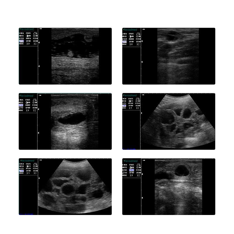
B-ultrasound or color ultrasound examination of the kidney can detect the following diseases: stones in the urinary system, which are manifested as high-echo areas with acoustic shadows behind them. In addition, water accumulation in the kidney can also be detected. There are also cystic spaces in the kidney, such as renal cysts, which are also relatively clear in B-ultrasound. In addition, solid spaces in the kidney, that is, renal cancer, are manifested as soft tissue spaces with blood flow in B-ultrasound. Congenital kidney malformations lead to narrowing and twisting of the junction of the renal pelvis and ureter, causing hydronephrosis and thinning of the renal cortex, which can all be detected through B-ultrasound. Yonkermed Medical is a B-ultrasound machine manufacturer. It has a variety of portable color ultrasound machines and cart-type B-ultrasound machines for use in hospitals, clinics, and veterinary medicine.
At Yonkermed, we pride ourselves on providing the best customer service. If there is a specific topic that you are interested in, would like to learn more about, or read about, please feel free to contact us!
If you would like to know the author, please click here
If you would like to contact us, please click here
Sincerely,
The Yonkermed Team
infoyonkermed@yonker.cn
https://www.yonkermed.com/
Post time: Sep-25-2024